Research Article - Journal of Natural Product and Plant Resources ( 2017) Volume 7, Issue 2
Phytochemical screening, antimicrobial and GC-MS analysis of the ethyl acetate and methanol crude extracts of the aerial parts of Palista hirsota and Raovofovia vomiti were studied in this work. The aim of the work is to ascertain the strength of the different solvent extracts of both plants in terms of microbial potency and chemical composition since both plants are used in Agbarho community for the treatment is bacteria and fungi infections. The preliminary screening of the various extracts was carried out using standard methods and the results revealed the presence of steroids and saponins for the ethyl acetate extract of Raovofovia vomiti while terpenes, steroids, flavonoids carbohydrate and saponins were present in the methanol extract of Palista hirsota. The antimicrobial screening was carried out on both plant extracts using the organisms; Salmonellae Typhi, Esherichia coli, Staphylococcus aureus, Klebsiella Pneumonia, Pseudomonas, Candida Krusei and Candida albican. The extract from Raovofovia vomiti produced a zone of inhibition of 20 mm, 10 mm and 10 mm for the organisms Salmonellae Typhi, Staphylococcus aureus, and Candida albican respectively with a corresponding MBC/MFC of 50mg/ml each while the extract from Palista hirsota has significant zone of inhibition on all the organisms tested against with a corresponding MBC/MFC >100mg/ml for organisms. The major compound detected by the library search of the data base in the GC-MS analysis revealed that both compound G1 and D1 from the aerial parts of the plants P. hirsota and R. vomitoria is octadecanoic acid also called stearic acid with the molecular formula C18H36O2, molecular weight 284g/mol and with a percent peak area of 45.58% for compound D1 from the aerial parts of R. vomitoria and 29.27% for Palista hirsota
Antimicrobial, Chemical compositions, GC-MS, Raovofovia vomiti, Organisms, Palista hirsota.
Bacteria and fungi diseases are caused by a pathogenic organisms present within the human environment. These diseases are common in most West African countries including Nigeria. It has been reported by WHO that approximately 60% of the inhabitants of most communities in the world believe in the use of medicinal herbs in the management of diseases. In many part of the world medicinal plant has been used for antibacterial, antifungal and antiviral activities for hundreds of years [1-3]. Rauvolfia vomitoria and Palisota hirsuta are common herbs used by the indigenes of Agbarho community of Delta state for the management of bacteria and fungi infections. It is against this background the chemical constituents of the different extracts of the aerial plant parts of these two plants are checked to ascertain chemical potencies of each plant and where possible proffer recommendations. Rauvolfia vomitoria and Palisota hirsuta are shrub or small tree found in different parts of West Africa including Nigeria. Recent studies have demonstrated the efficacy of different the Rauvolfia species used extensively for various ailments. It is useful in the lowering of blood pressure [4]. Palisota hirsuta are used in Ghana and other West African states for various painful and inflammatory conditions. The anti-inflammatory conditions of the ethanolic leaf extract of Palisota hirsuta was assessed and reported by woode Ogwuche and Adeyemi 2016, investigated the in vitro antimicrobial activity of the methanol extract of R. vomitoria used in Agbarho community of Delta state and it revealed that the extract was bactericidal against the following organisms, Staphylococus aureus, Klebsiella pneumonia, Pseudomonas, Candida albicans and Candida krusei at different concentrations and also other chemical constituents were also ascertained.
Plant material
The aerial part of the plants Palisota hirsuta and Rauwolfia vomitoria were collected from Agbarho community and identified at the herbarium of University of Benin. They were dried in the laboratory at ambient temperature of around 25°C; plant materials were individually crushed using a mortar and pestle to provide a greater surface area. The crushed plant materials were weighed and it gave 176 g for Rauwolfia vomitoria and 157 g for Palisota hirsuta, thereafter they were placed in container which were labelled and kept at room temperature. Crude plant extract was obtained by Soxhlet extraction method.
Phytochemical screening
The extracts of both plants were obtained differently using Soxhlet extractor and they were subjected to phytochemical screening using standard techniques of plant secondary metabolites by Harborne [5], Sofowora [6] and Trease and Evans [7]. The crude plant extracts of the aerial plants of both plants were tested for Alkaloids, Saponins, Phlobotannins, Phytosterolds Terpernoid, Phenols, Carbohydrate, Tannins, Steroids, Flavonoids and Cardiac Glycosides.
Chromatography
Thin layer chromatography (TLC) was conducted on Silica gel (E-Merck and BDH) coated on a thin glass plate to determine the solvent combination to use for column chromatography and to view other components. Spots on TLC were detected by viewing under florescent lamp and further by spraying with 20% tetraoxosulphate (VI) acid, followed by heating at 60°C. Column chromatography was carried out on the extract over silica gel using gradient elution method with different solvent systems in order of increasing polarity.
Methanol extract
A combination of methanol and ethyl acetate solvent was used to elude for the methanol extract. 5 g of the methanol extract was loaded on the column. 100% ethyl acetate was initially used to run the column. Thereafter, by using gradient elution, the subsequent ratios used were 8:2, 7:3, 6:4, 5:5 up to 100% methanol and a total of ten fractions were collected at each solvent mixture. These fractions were allowed to evaporate to dryness. Another Thin layer chromatography (TLC) was conducted on the fractions collected, thereafter similar fractions were pooled together and the purer crystal was taken for analysis labeled, G1.
Ethyl acetate extract.
A combination of petroleum ether and ethyl acetate solvent was used to elude the ethyl acetate extract. 5 g of the ethyl acetate extract was loaded on the column. 100% petroleum ether was initially used to run the column. Thereafter, by using gradient elution, the subsequent ratios used were 8:2, 7:3, 6:4, 5:5 up to 100% ethyl acetate and the column was finally washed with methanol and a total of ten fractions were collected at each solvent mixture. These fractions were allowed to evaporate to dryness. Another Thin layer chromatography (TLC) was conducted on the fractions collected, thereafter similar fractions were pooled together and the purer crystal was taken for analysis labeled, G1.
A combination of petroleum ether to ethyl acetate solvent was used. At a ratio of 8:2, seven fractions were collected, at the ratio of 7:3, ten fractions was collected and at the ratio of 6:4 five fractions was collected. The column was finally washed with methanol (50 ml). These fractions were allowed to evaporate to dryness. Thin layer chromatography (TLC) was conducted on the fractions collected, thereafter similar fractions were pooled together and the purer crystal was taken for analysis labeled, D1.
Antimicrobial Screening
Zone of inhibition
The antimicrobial activities of the extracts from both plants were determined using some pathogenic microorganisms. The test microbes such as Salmonellae Typhi, Esherichia coli, Staphylococcus aureus, Klebsiella Pneumonia, Pseudomonas, Candida Krusei and Candida albican were obtained from Emma-Maria Biometric laboratory Abraka, Delta State. The zone of inhibition was conducted using the method of Kumara et al., [8].
Culture media
The culture media used were Mueller Hinton agar (MHA) and Mueller Hinton broth (MHB). All the media were prepared according to manufacturer's specifications.
Preparation of inoculums of test organisms
The McFarland turbidity standard scale 1 was used to standardize the organisms. The scale was prepared by adding 9.9 ml of 1 % barium chloride (BaCl2). Suspensions of the organisms were made in sterile distilled water and compared with the McFarland turbidity standard, until the opacity match with the scale number 1, which corresponds to 1.5 x 10⁶ CFU/ml.
Determination of MIC of plant extract by micro dilution method
Test-tubes were prepared by dispensing 50 μl of Nutrient broth for bacteria, into each well. A 50 μl from the stock solution of tested extract (concentration of 200 mg/ml) was added into the first row of the plate. Then, twofold, serial dilutions were performed by using a micropipette. The obtained concentration range was from 100 to 25 mg/ml, and then added 10 μl of inocula to each test-tube except a positive control (inocula were adjusted to contain approximately 1.5 x 10⁸ CFU/mL). The extracts of both plants were individually used, with the media as a positive control and inoculum with media was used as a negative control. The test plates were incubated at 37°C for 18 hours [9,10].
Gas chromatography and mass spectrometry (GC-MS)
GC-MS analysis was carried out on each extract. It was analyzed using GC-MS QP2010 Plus Shimadzu under the following condition: column used were Rtx-5MS, 30 m length and inner diameter of 0.25 mm and the initial column temperature was 80°C and final temperature was 280°C, while the injector temperature was 250°C with split mode injector and split ratio of 1 and pressure of 108.0 kPa. The flow rate was 6.2 ml/minute and the flow within the column was 1.58 ml/minute. The detector temperature was 230°C and using Helium as the gas carrier with FID (Flame ionization detector); and the samples volume injected was 8 μl. Compounds were identified by comparing retention indices/comparing mass spectra of each compound with those of authentic samples and library
FTIR-84005 fourier transform infrared spectrophotometer
The Infra-red spectra's were recorded on FTIR-8400S (Shimadzu Deutchland GmbH) spectrophotometer in KBr and polyethylene pellets. The extract was weigh-in at 0.01 g and homogenized with 0.01 g KBr anhydrous by mortar agate. The mixture of sample and KBr were pressed by vacuum hydraulic at 1.2 psi (pounds per square inch) to obtained transparency pellet. Samples were usually scanned in the absorption area of 500-4000 cm-1. The results of analysis consisted of chemical structure, molecular binding form and certain functional group of tested sample as basic of spectrum type.
Phytochemical screening
The phytochemical screenings of the extracts of plants are shown in Table 1. It revealed the presence of Steroid, cardiac glycosides, phytosterol, terpenoid. Researchers have reported that some secondary metabolites can block tumour growth in rodent models, which further supports the idea that they have potential for cancer therapy and they also have pharmacological activities which include antihypertensive effect, anti-malarial activities and anticancer actions [11,12] (Tables 2-5).
| Constituents | Test | Observation | Inference | |
|---|---|---|---|---|
| Ethyl RAÂ Â | ME PA | |||
| Alkaloid | Extract + 2ml 1% HCl. Filtrate + Wagner reagent | Formation of yellow colouration, turn reddish brown on addition of Wagner reagent | Absent | Absent |
| Terpernoid | Extract + 2ml Chloroform +3ml of conc. H2SO4 | Formation of yellow colouration, turn green on addition of conc. H2SO4 | Absent | Present |
| Phenols | Extract + 4 drops of FeCl3 | Green colouration | Absent | Absent |
| Steroid | Extract + 2ml chloroform + 3 drops conc. H2SO4 | Yellow colour, on addition of conc. H2SO4 turn green | Present | Absent |
| Flavonoid | Extract + 2ml of 2% NaOH | Green colouration, turn light green | Absent | Present |
| Phytosterol | .Extract + 2ml Chloroform. Filtrate + 3 drops of conc. H2SO4 | Yellow colouration, turn green on addition of conc. H2SO4 | Absent | Absent |
| Carbohydrate | Extract + 2ml Bennedict solution + heat | Blue colouration. on heating turn green | Absent | Present |
| Phlobatanin | Extract + 1% HCl | Light green colouration | Absent | Absent |
| Saponin | Extract + 20ml distilled H2O, shake for 15 minute | Colourless | present | Present |
| Tannin | Extract + 10ml distilled H2O. Filtrate + 2ml FeCl3 | Colourless and turn yellow on addition of FeCl3 | Â Absent | Absent |
Table 1: Qualitative phytochemical screening of Rauvolfia vomitoria and palista histrus.
| Organisms | EAc RA(mm) | ME PA(mm) |
|---|---|---|
| Staphylococcus aureus | 12 | 4 |
| Escherichia coli | 0 | 16 |
| Salmonellae Typhi | 10 | 28 |
| Klebsiella pneumoniae | 0 | 15 |
| Pseudomonas aeruginosa | 0 | 15 |
| Candida albicans | 10 | 18 |
| Candida krusei | 0 | 14 |
| Key: 0 = No zone of inhibition, EAc RA = Ethylacetate extract of Raufofovia vomite, M.E = Methanol extract of Palista histrus. | ||
Table 2: Comparison of the zone of Inhibition (sensitivity test) for Rauvolfia vomitoria extract and palista histrus extracts .
| Organisms | AU | CPX | PN | CEP | OFX | NA | PEF | CN |
|---|---|---|---|---|---|---|---|---|
| Staphylococcus aureus | 15 | 0 | 10 | 10 | 10 | 0 | 0 | 11 |
| Escherichia coli | 16 | 0 | 0 | 13 | 16 | 0 | 0 | 0 |
| Salmonellae Typhi | 10 | 0 | 0 | 12 | 18 | 0 | 0 | 10 |
| Klebsiella pneumonia | 12 | 0 | 0 | 12 | 0 | 0 | 0 | 0 |
| Pseudomonas aeruginosa | 15 | 0 | 0 | 10 | 0 | 0 | 0 | 0 |
| Key: AU-AUGUMETIN, CPX-CIPROFLOX, PN- AMPLICIN, CEP-CEPOREX, NA-NALIDIXIC ACID, PEF-REFLACINE, OFX-TARIVID, CN-GENTAMYCIN | ||||||||
Table 3:Â Zone of Inhibition (sensitivity test) for the antibiotic disc (drugs) Extract in diameter (mm).
| ORGANISMS | Nystatin 1000UI | Fluconazole 230mg |
|---|---|---|
| Candida albicans | 0 | 12 |
| Candida krusei | 10 | 6 |
Table 4: Zone of Inhibition (sensitivity test) for the antifungal disc (drugs) Extract in diameter (mm).
| Organisms | 100mg/ml | 50mg/ml | 25mg/ml | 12.5mg/ml | 6.25mg/ml | MBC |
|---|---|---|---|---|---|---|
| Methanol extract PA | ||||||
| Staphylococcus aureus | ++- | +-- | --- | --- | --- | 50mg/ml |
| Escherichia coli | +++ | +-- | --- | --- | --- | 50mg/ml |
| Salmonellae Typhi | +++ | ++- | --- | --- | --- | 50mg/ml |
| Klebsiella pneumonia | +++ | ++- | --- | --- | --- | 50mg/ml |
| Pseudomonas aeruginosa | +++ | ++- | --- | --- | --- | 50mg/ml |
| Ethyl Acetate extract RA | ||||||
| Staphylococcus aureus | --- | --- | --- | --- | --- | >100mg/ml |
| Escherichia coli | --- | --- | --- | --- | --- | >100mg/ml |
| Salmonellae Typhi | --- | --- | --- | --- | --- | >100mg/ml |
| Klebsiella pneumonia | --- | --- | --- | --- | --- | >100mg/ml |
| Pseudomonas aeruginosa | --- | --- | --- | --- | --- | >100mg/ml |
| Key: ++=No inhibition on both runs, +- =Inhibition on one run and no inhibition on the second run, - - = Inhibition on both runs, PA= Palista histrus RA = Raufofovia vomite | ||||||
Table 5: Comparison of the Minimum Inhibitory Concentration of Rauvolfia vomitoria extract and Palista histrus extracts on some selected bacteria isolates.
| Bands (cm-1) | Functional group |
|---|---|
| 3443.05 | O-H Stretch |
| 2935.76 | C-H Stretch of alkanes |
| 1647.26 | C=C Stretch of alkenes |
| 1425.44 | -CH3 bend f alkanes |
| 1051.24 | C-O Stretch present in compounds with C-O bonds ethers, esters etc. |
Table 6: Functional groups in Compound G1 analyzed by using FTIR.
| Bands (cm-1) | Functional group |
|---|---|
| 2927.08 | O-H Stretch |
| 1646.30-1540. 21 | N-H bend of primary and secondary amine |
| Â 1421.50 | C-H Stretch of a -CH2- bend of |
| 1078. 24 | C-O Stretch of esters, ethers, carboxylic acids and anhydride |
Table 7: Functional groups present in compound D1 analyzed by using FTIR.
| Peak Number | Retention Time 3 | Area% | Height% | Formula | Molecular Weight | Compound Name |
|---|---|---|---|---|---|---|
| 1 | 3.525 | 0.24 | 0.67 | C6H14O2 | 118 | 1-Propanol, 2-(1-methylethoxy)- |
| 2 | 4.300 | 1.45 | 1.18 | C8H16 | 112 | 2-Octene |
| 3 | 5.967 | 5.29 | 4.64 | C5H10O4 | 134 | 1,2,3-Propanetriol |
| 4 | 7.533 | 1.46 | 3.36 | C5H10O4 | 134 | 1,2,3-Propanetriol |
| 5 | 9.558 | 3.55 | 2.17 | C4H8O3 | 104 | 1,2-Ethanediol, monoacetate |
| 6 | 10.775 | 1.93 | 2.12 | C8H12O6Â Â | 204 | 1,1,2-Triacetoxyethane |
| 7 | 13.142 | 6.87 | 3.06 | C6H13ClO2Â | 152 | 2-Propanol, 1-chloro-3-propoxy |
| 8 | 13.958 | 7.46 | 4.13 | C4H8O3Â | 104 | 1,2- Ethanediol monoacetate |
| 9 | 15.067 | 2.00 | 1.93 | C11H20O2 | 184 | 3,6-Dimethyl-5-hepten-1-ol acetate |
| 10 | 15.225 | 0.85 | 1.82 | C14H28OÂ | 212 | E-2-Tetradecen-1-ol |
| 11 | 16.808 | 1.81 | 3.46 | C17H34O2 | 270 | Hexadecanoic acid, methyl ester |
| 12 | 18.042 | 18.95 | 17.50 | C15H30O2 | 242 | Pentadecanoic acid |
| 13 | 19.933 | 1.20 | 2.81 | C19H36O2 | 296 | 6-Octadecenoic acid, methyl ester |
| 14 | 20.192 | 3.65 | 8.00 | C20H40O | 296 | Phytol $$ 2-Hexadecen-1-ol, 3,7,11,15-tetramethyl-, |
| 15 | 20.308 | 0.67 | 1.55 | C22H44O2 | 340 | Heneicosanoic acid, methyl ester |
| 16 | 20.783 | 9.95 | 13.38 | C16H30O2Â | 254 | Hexadecenoic acid, |
| 17 | 21.058 | 29.27 | 22.84 | C18H36O2Â | 284 | Octadecanoic acid OR Stearic acid |
| 18 | 21.517 | 1.63 | 3.22 | C17H32O2Â | 268 | Heptadecenoic acid |
| 19 | 23.325 | 23.184Â | 0.68 | C19H38O2 | 298 | Nonadecanoic acid |
| 20 | 26.450 | 1.07 | 1.01 | C21H40O3Â | 340 | Oleic acid, 3-hydroxypropyl ester |
Table 8: Sample C (Palista hirsota).
Spectroscopic measurement
The both compounds G1 from Palista crude extract and D1 from Rauvofovia crude extracts were analyzed using FTIR-8400S Fourier Transformer Infrared Spectrophometer and an Agilent Gas Chromatography (6890N model) coupled to 5973 Mass Selective detector (MSD), using chloroform as solvent at the National Research Institute of Chemical Technology Zaria. The results obtained were compared with an inbuilt main library C:/Database/NIST02.L) and this library enabled the confirmation of the compound(s) present in the plant (Figures 1, 2 and Tables 6-9).
| Peak Number | Retention Time 3 | Area% | Height% | Formula | Molecular Weight | Compound Name |
|---|---|---|---|---|---|---|
| 1 | 3.508 | 0.72 | 1.69 | C8H18O | 130 | 2-Heptanol, 3-methyl |
| 2 | 4.125 | 1.93 | 2.95 | C7H14 | 98 | 1-Heptene |
| 3 | 6.108 | 15.36 | 10.46 | C7H14O2Â | 130 | 1-Butanol, 3-methyl-, acetate |
| 4 | 16.817 | 3.11 | 5.16 | C17H34O2Â Â | 270 | Pentadecanoic acid, 14-methyl-, methyl ester |
| 5 | 17.883 | 17.64 | 18.57 | C16H30O2Â | 254 | Hexadecenoic acid, |
| 6 | 19.825 | 0.99 | 2.18 | C12H20 , |
164 | 1,6,11-Dodecatriene |
| 7 | 19.925 | 2.78 | 6.01 | C19H36O2Â | 296 | 9-Octadecenoic acid, methyl ester |
| 8 | 20.167 | 1.20 | 2.53 | C20H40O | 296 | Phytol or 2-Hexadecen-1-ol, 3,7,11,15-tetramethyl |
| 9 | 20.292 | 45.58 | 33.36 | C18H36O2Â | 284 | Octadecanoic acid OR Stearic acid |
| 10 | 20.833 | 1.35 | 3.01 | C22H42O2Â | 338 | Erucic acid or Docosenoic acid |
| 11 | 21.042 | 1 9.34 | 14.09 | C16H30O2Â | 254 | Hexadecenoic acid |
Table 9: Sample B (Rauvolfia vomitoria).
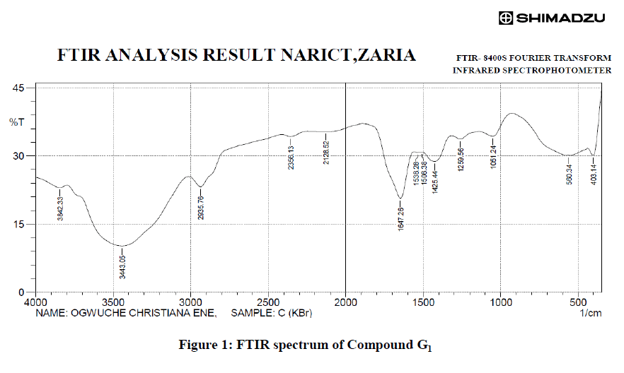
Figure 1: FTIR spectrum of Compound G1
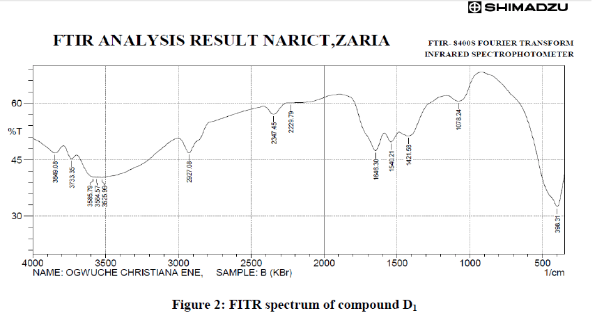
Figure 2: FITR spectrum of compound D1.
GC-MS analysis of compound G1 and D1
GC-MS analysis was carried out on Agilent Technologies 6890N Network GC System and Agilent Technologies 5973 Network Mass Selective Detector coupled with 7683B Series Injector. The model number of the column used was Agilent 122-5533 capillary column with specification: DB-5 ms, 0.25 mm*30 m*1 μm. The carrier gas used was Helium at a flow rate of 1.2 ml/min. The injection volume was 1 μ. The inlet temperature was maintained at 230°C. The oven temperature was programmed initially at 5°C for 5 minutes, Then programmed to increase to 300°C at a rate of 10°C ending with 25 minutes. Total run time was 45 minutes. The MS transfer line was maintained at a temperature of 300°C. The source temperature was maintained at 230°C and the MS Quad at 150°C. The ionization mode used was electron ionization mode at 70 eV. Total Ion Count (TIC) was used to evaluate for compound identification and quantitation. The Spectrum of the separated compound was compared with the database of the spectrum of known compound saved in the NIST02 Reference Spectra Library. Data analysis and peak area measurement was carried out using Agilent Chemstation Software (Figure 3).
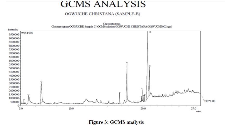
Figure 3: GCMS analysis.
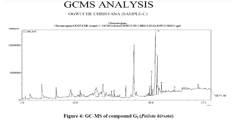
Figure 4: GC-MS of compound G1 (Palista hirsota).
Identification of components
The database of National Institute of Standard and Technology (NIST) having more than 62,000 patterns was used to identify the compound. The spectrum of the unknown components was compared with the spectrum of the known compounds stored in the NIST library (Figures 4 and 5).
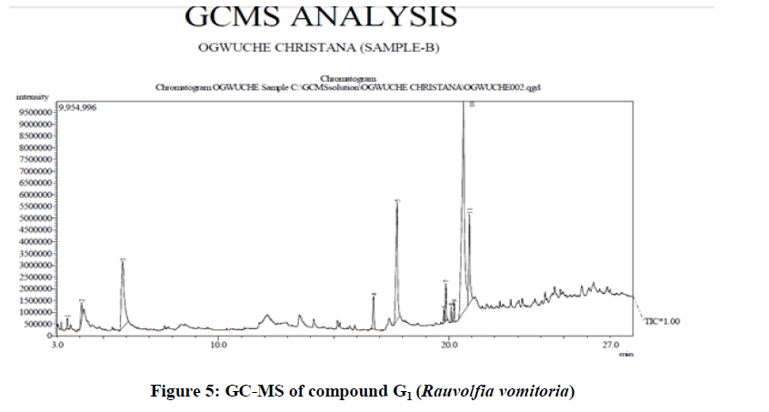
Figure 5: GC-MS of compound G1 (Rauvolfia vomitoria).
The major compound detected by the library search of the data base in the GC-MS analysis revealed that both compound G1 and D1 from the aerial parts of the plants Palista hirsota and Rauvolfia vomitoria respectively is octadecanoic acid also called stearic acid with the molecular formula C18H36O2, molecular weight 284 g/mol and with a percent peak area of 45.58% for compound D1 from the aerial parts of R. vomitoria and 29.27% for Palista hirsota. Stearic Acid is a saturated long-chain fatty acid with an 18-carbon backbone. Stearic acid is found in various animal and plant fats.
Stearic acid It has been reported to have antibacterial, antifungi, antiviral and anti-inflammatory activities, as a result, cream formulations containing n-docosanol (docosanol) or stearic acid were tested for effects on chemically-induced burns in mice [13].
Also from the hexane-soluble fraction of an ethanol extract from leaves and stems of Stemodia foliosa (Scrophulariaceae), the new stearic acid 4-[(n-pentoxy) phenethyl] ester (1) was isolated. This compound exhibited antibacterial properties at 10 microg/mL concentration by using disc diffusion method against Gram-positive bacteria Bacillus cereus and Bacillus subtilis and fast-acid bacterium Mycobacterium fortuitum [14].
Manivachagam, et al., [15], reported the gas chromatographic analysis of fatty acid methyl esters from Sesuvium (S.) portulacastrum antimicrobial activity against human pathogenic microorganisms. The analysis revealed the presence of palmitic acid with the highest relative percentage (31.18%), followed by oleic acid (21.15%), linolenic acid (14.18%) linoleic acid (10.63%), myristic acid (6.91%) and behenic acid (2.42%). The saturated fatty acids were higher than the unsaturated fatty acids. The result showed the highest antibacterial and anticandidal activities and moderate antifungal activity against the tested microorganisms Bacillus subtilis, Aspergillus fumigatus and Aspergillus niger at different zones of inhibition [16].
This study illustrates that the extracts of the aerial parts of Palista hirsota and Raovofovia vomiti used for the management of bacterial and fungi diseases in Abgarho community of Delta state are good sources of metabolites with antimicrobial activities worthy of further investigations. The extracts were bactericidal against the causative organism Salmonella typhi in which they are mostly used for the treatment of typhoid fever and also against other organisms tested against such as Staphylococcus aureus, Escherichia coli, Pseudomonas aeruginosa, Klebsiella pneumoniae Candida albicans and Candida krusei at different MIC and MBC/MFCs.
Authors wish to thank Federal University of Petroleum Resources Effurun-Nigeria, for providing an enabling laboratory environment for this research work. We wish to thank Professor M.O. Edema for taking the plant, all the way, to the Herbarium of the University of Benin - Nigeria for Identification.