Research Article - European Journal of Sports & Exercise Science ( 2020) Volume 8, Issue 1
Received: 01-Apr-2020 Published: 17-Apr-2020
Context: Photobiomodulation (PBM) has been used to treat musculoskeletal injuries, reduce pain, heal tissue, and improve muscle performance and function. Many physiological effects of PBM therapy have been examined, but the thermal effects of PBM therapy have yet to be determined. Objective: To determine the superficial tissue heating characteristics of a Red and Blue combination light patch. Design: Controlled laboratory study. Setting: Research laboratory. Patients or Other Participants: Ten healthy individuals (M=5, F=5, age 22.2 ± 2.3, height (170.3 ± 13.1 kg). Interventions: Participants were positioned prone on a padded treatment table. While lying on the table, their posterior calf was cleansed before an IT-21 needle thermocouple was inserted 0.5 cm into the subcutaneous tissue. A PT-IT needle was secured to the posterior calf adjacent to the needle thermocouple. A second PT-6 thermocouple was secured to the skin 3 cm away from the treatment location. All thermocouples interfaced with an isothermex electrothermomitor. A single Careware Firefly (Carewear, Reno, NV) light patch was positioned over the subcutaneous and skin thermocouple within the treatment area. A 15 minute PBM therapy (wavelength=640 and 450 nm, average irradiance=3 m/W/cm2, peak power=9 mW, continuous peak power=3 mW, energy density=5.4 J/cm2, treatment area=50 cm2) was administered to the posterior calf. After the treatment, a 5-minute post-treatment temperature decay was recorded. Main Outcome Measures: Tissue temperature measurements from the thermocouples were taken every 1 minute throughout the 15-minute treatment and for 5 minutes post-treatment. Absolute tissue temperature and change from baseline temperature were used during the data analysis. A repeated measures ANOVA was used to determine if tissue temperature increased between the 3 measurement sites over the treatment time. Results and Conclusion: The red and blue wavelength combination photobiomodulation light patch significantly increases superficial tissue temperature. Thermal physiological effects may be an added benefit to this type of PBM therapy.
At the October 2018 World Association of Laser Therapy (Nice, France) the term laser was changed to Photobiomodulation. PMB therapy has shown very little temperature increase in healthy normals. High Power, Class IV lasers have produced a vigorous temperature increase of 4-5°C. We wanted to see if low power lasers would increase the temperature in healthy normals, using a novel blue (450 nm) and red (645 nm) LED PBM therapy patch. This has never been tested with low power lasers.
Photobiomodulation is the use of non-ionized light sources, including lasers and Light Emitting Diodes (LEDs), in the visible and near infrared spectrum. PBM stimulates endogenous chromophores eliciting various biological events within the tissues [1]. The biological events are largely dependent on the PBM therapy’s wavelength, which determine the therapy’s specific color of light [2]. Different wavelengths in the visible, from blue to red, and infrared spectrums have been used in treating musculoskeletal conditions in physical medicine and rehabilitation.
Participants
We recruited and enrolled 10 healthy males (N=5) and females (N=5) between the age of 18-35 years old. Participants had to refrain from performing lower extremity strength training at least 4 times, 1 month prior to enrollment and if the participant presented with contraindications or precaution related to photobiomodulation, such as cancer, recent organ transplant, epilepsy, had a steroid injection within the past 3 months, an acute infection, and/or suffers from a thyroid condition.
Before participants were enrolled, the study procedures were approved with the Institutional Review Board at Brigham Young University. All participants provided written informed consent before all procedures were performed.
Therapeutic intervention
Participants were randomly assigned to the active or sham treatment group. Those who received the sham group would have the treatment given to them the next day. The subjects who started with the treatment group who were treated with the sham group the next day. The sham treatment was performed by turning on the devices power control but a switch built into the device allowed the investigator to turn off the power to the light patch. The participant was instructed during the sham treatment that an infrared treatment was being applied.
Photobiomodulation therapy was delivered with 2 LED light patches (Model: Firefly, Carewear Corp, Reno, NV) applied to the posterior aspect of the triceps surae (calf) muscle. A 25 gauge needle thermistor was inserted 1.5 cm deep from the skin’s surface. The other end was inserted into the Isothermix that records temperature change. The 50 cm2 light patch incorporates blue and red light (wavelengths: 640 and 450 nm) with an average irradiance of 3 mW/cm2. On each light patch, there are 130 hexagon shaped clusters of multiple micro LEDs. The LED PBM therapy was continuous (a 100% duty cycle). The average W was 9 mW/cm2. The ratio of blue to red optical power was 3:1. We delivered the PBM therapy for 15 minutes creating a fluence of 5.4 J/cm2. When using 2 light patches the total treatment area was 100 cm2. The LED light patch set-up is visualized in Figures 1-3.
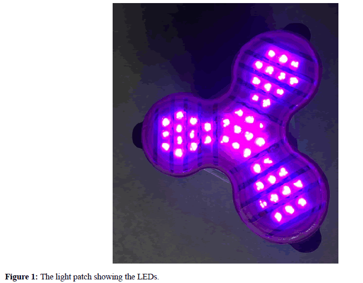
Figure 1: The light patch showing the LEDs.
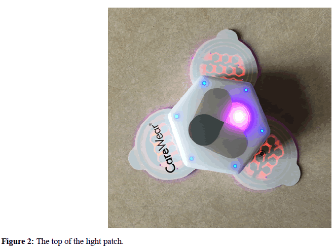
Figure 2: The top of the light patch.
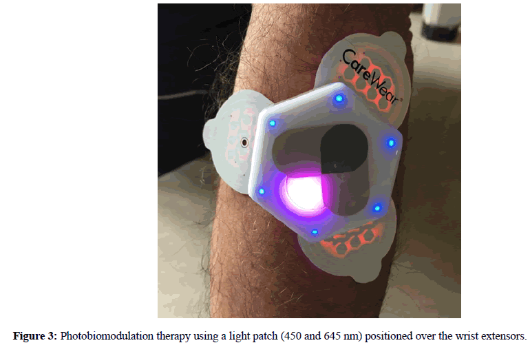
Figure 3: Photobiomodulation therapy using a light patch (450 and 645 nm) positioned over the wrist extensors.
The light patch increased subcutaneous tissue temperature 6.22 ± 2.25°C to a peak absolute temperature of 37.71 ± 1.78°C. The light patch increased tissue temperature 8.22 ± 2.62°C to a peak absolute temperature of 38.90 ± 2.24°C. The skin temperature 3 cm away from the treatment remained relatively constant throughout the treatment (-0.11 ± 0.30°C). The subcutaneous and skin temperatures directly under the treatment area significantly heated compared to the reference 3 cm away from the treatment site (p<0.001). During the 5 minutes post-treatment, tissue temperatures decreased 2.68 ± 1.45°C, 4.54 ± 1.93°C, and 0.23 ± 0.12°C at the subcutaneous treatment skin surface, and 3 cm away from skin surface sites, respectively.
The simple to use, novel, wearable, active blue-red LED light patch created a positive outcome in the actual treatment. There was no or very little increase in the sham treatment group. Blue light has traditionally been known for its bactericidal effects, but new research has highlighted its effects for other tissue types. First, blue light stimulates light-gated ion channels known as opsins, which have a role in tissue calcium release. Calcium has a vital function during muscle function and wound healing. Second, blue light also stimulates flavins, which are biological chromophores that repair damaged DNA. Finally, blue light stimulates nitric oxide release in mitochondria subject to inflammation, restoring mitochondrial respiration and increasing local vasodilation [3-5].
Red light has been a primary waveform for the treatment of musculoskeletal conditions because of its known effects on mitochondrial ATP production. Red light stimulates unit IV, cytochrome C oxidase, in the electron transport chain leading to increased enzyme activity, oxygen consumption, and ultimately greater ATP production [2]. Recently, it has also been established that red light causes dissociation of bound nitric oxide in the mitochondria, found when the tissues are in an oxidative stressed environment. The dissociation of nitric oxide leads to less reactive oxidative species and less tissue damage when in a stressed state [3,4]. Near-infrared wavelengths have similar functions to red light, with better tissue penetration [6-10].